मधुमक्खियों की 9 प्रमुख बीमारियॉं तथा उनसे बचाव के तरीके
Beekeeping is an interesting hobby, an ideal agro-based subsidiary enterprise, providing supplementary and sometimes major income to the people in the rural areas. The diverse fauna of honeybees occurring in India further enhances its practical utility.
Although seven species of Apis have been described but India is a unique country where the four Apis species of major importance are now present. These include two wild species viz. Apis dorsata (Giant / Rock honey bee or dumna) & A. florea (small honey bee) and two domesticated species viz. A. cerana (oriental honey bee) and A. mellifera (occidental or European honey bee).
The first three species are indigenous, while the fourth one is an exotic introduction in the country. Like all other living creatures, honey bees also suffer from several diseases and are attacked by different enemies..
The diseases in brood and adult honey bees are caused by bacteria, fungi, viruses, rickettsiae and protozoa. In India, brood diseases such as American foul brood, European foul brood, Thai sac brood and adult bee diseases viz. acarine, nosema and clustering disease have been reported in Asiatic hive bee, Apis cerana Feb.
European honey bee, Apis mellifera Linnaeus have been reported to suffer from European foul brood, sac brood and chalk brood diseases. Bees have two distinct life forms (brood and adult) and most diseases are specific to either one stage or the other but the most virulent diseases are those of the brood.
A brief account of various diseases and their management is given below:
I. Brood Diseases:
They are generally easier to recognize as a group than adult diseases but are more difficult to control. These are either caused by bacteria or fungi or virus.
a) Bacterial disease
1) American Foulbrood (AFB)
It is caused by spore forming bacterium, Paenibacillus larvae.In temperate and sub-tropical regions it is the most virulent brood disease as it forms the heat and drought-resistant spores which are capable of surviving for more than 35 years and germinate as and when get the favorable environment.
In tropical Asia, where sunlight is abundant and temperatures are relatively high throughout the year, the disease seldom causes severe damage. Death of an infected larva takes place after the cell has been sealed and the cocoon has been spun.
Symptoms:
- At death, the diseased larva changes from a normal pearly white color to a creamy brown, then gradually darkens.
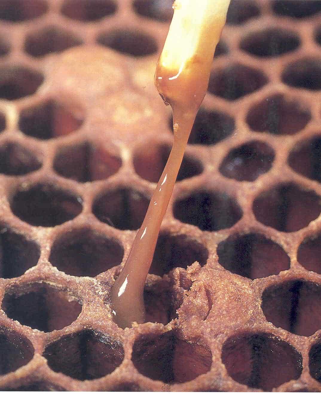
- When a match-stick is thrust into the cell of the decomposed pupa, it draws out a ropy thread of several centimeters in length (Fig. 1).
- As the larva dries up, it becomes dark brown or black, rather rough scale that lies uniformly on the lower side of the cell. These scales stick very tightly to the cell wall and can be removed only with great difficulty.
- The decomposed brood has an unpleasant smell.
- The normal convex cell cap becomes moist, dark and sunken, and later perforate. The perforation of the capped cells is the result of the attempt by the workers to uncap it to remove the decomposing remains.
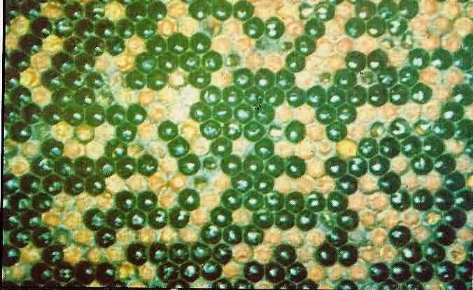
- The brood combs of an affected colony become patchy in appearance (Fig. 2), owing to the presence of the intermixed diseased and healthy
2) European Foulbrood (EFB)
It is caused by the non-spore-forming bacterium, Melissococcus plutonius, which invades the mid-gut of 4-5 days old larvae and multiplies rapidly causing death.This is a disease of unsealed (open) brood as the worker bees may leave the cell containing the dead larva uncapped. Sometimes the infected larva does not die until it is sealed, and this may result in sunken and perforated cappings.
Symptoms:
- Infected larvae move about inside the cell instead of staying in the normal curled position as a result, the dead larva is found in an unnatural coiled position across the mouth of its cell.
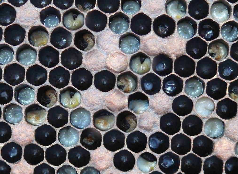
Dead larvae become soft, watery and dull yellow. Their breathing tubes are prominent at this watery stage. The affected larvae are discoloured, first creamy yellow, then turn to light brown and then dark brown and occasionally black.
- The infected larvae lie upright attached with side walls of the cells and sometimes appear just melted down at the base of the cells.
- Dead larvae finally dry to brown removable rubbery scales at the bottom of the cell. Brood pattern becomes irregular (Fig. 3).
- The bacteria may not cause any odor in infected colonies. However, secondary invasion by other bacteria could result in a sour or foul smell.
Spread of Bacterial Diseases
Within the colonies, these diseases are spread by nurse bees through their feeding activities. Besides the inoculum is also spread in the colonies by in house/ house cleaning bees which pick up the inoculum during cleaning off the infected larvae from the colonies. However, due to following reasons, the diseases are spread through:
- Inter colony substitution of bees, brood, queen bee and pollen or food reserves from infected colony to healthy colony.
- Robbing among the colonies.
- Drifting of the bees among the colonies in the apiary.
- Use of contaminated equipments in the healthy colonies.
- Hiving of swarms of unknown origin in one’s apiary may be source of inoculum of diseases for their spread to the other healthy colonies.
- Drifting of bees of adjacent apiaries or through the infected foragers of the adjacent apiary by leaving inoculum on the crop flowers which are also visited by bees from healthier colonies of other apiary.
Management:
- Sterilize the combs and other hive parts with Formalin @ 150 ml/ l water, for 48 h at 43oC in fumigation chambers.
- Sterilize the combs with ethylene oxide @ 1 g/l for 48 h at 43°C in fumigation chambers
- Breeding disease resistant strains of bees is one of the best measures for the disease management.
- Burning of colonies including swarm shook coupled with provisioning of either brood alone or brood + pollen combs from the healthy colony is effective in controlling the disease. This method is commonly followed in European countries.
b) Fungal Diseases
Two fungal diseases are important viz. Chalk brood and stone brood.
3. Chalk Brood
This disease is caused by spore-forming fungus, Ascosphaera apis. The spores of fungus remain viable for years. The disease is most prevalent in the spring when the brood area is expanding, and the weather is still cool and there are not enough nurse bees to maintain the brood nest temperature. Its endemic infection is damaging otherwise it is a less serious disease.
It affects only the brood. Brood cells can be sealed or unsealed. Workers, drones, and queens are all susceptible to the disease. Three - four days old larvae and those on periphery of brood area are more susceptible.
Symptoms:
- Diseased larvae are stretched out in their cells in an upright position. Larvae dead from chalk brood disease are chalk-white and are often covered with cottony filaments, hence the name "chalk brood" (Fig. 4).
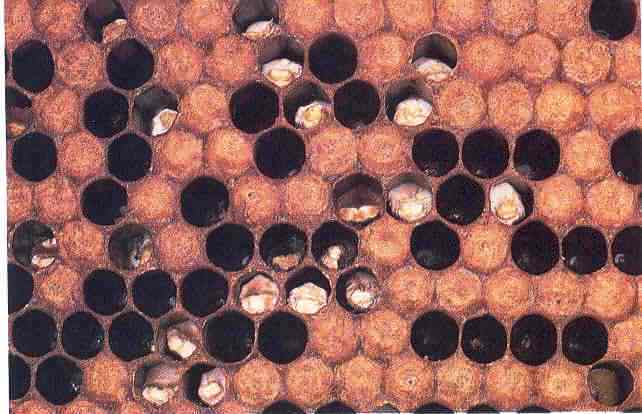
Sometimes the diseased larvae can be mottled with brown or black spots, especially on the ventral sides. The white coloration may eventually give way to a gray or black, depending on the life stage of the fungus.
- Chalk brood mummies once dry, are loose in the cell, and can be removed easily. Often, a few of these mummies are visible on the ground at the entrance to the hive.
- The disease is transmitted by spores that are readily moved from colony to colony on infected pollen, robbing bees, drifting bees, or beekeeping equipment.
4. Stone Brood
This disease is caused by Aspergillus flavus whichcauses mummification of the brood of a honey bee colony. The fungi are common soil inhabitants and are also pathogenic to other insects, birds and mammals.
Symptoms:
- Its spores are ingested with food and germinate in the gut, growing rapidly to form a collar like ring near the head.
- After death the larvae turn black and become difficult to crush, hence the name “stone brood”.
- Eventually the fungus erupts from the integument of the larva and forms a false skin.
- The affected adult bees show restlessness, feebleness and paralysis, abdomen gets dilated and then mummified.
- Younger bees die earlier.
Management of Fungal Diseases
- There is no chemical control.
- Removal of mummies by bees results in natural control of the diseases.
- Collect and burn the mummified larvae.
- Improve ventilation and reduce humidity.
- Replace old, blackened brood combs as these may harbor chalkbrood spores.
- If a colony lacks sufficient food stores, supplement with good-quality feed.
- Replace queens with stock bred for hygienic behavior and/or disease resistance.
c) Viral Diseases
5. Sac Brood Virus (SBV) and Thai Sac Brood Virus (TSBVS)
TSBV is a virulent strain of SBV of Apis mellifera.Disease symptoms for diagnosis of both the diseases are similar. While TBV is infective on Apis cerana, TSBV infects Apis mellifera.
Symptoms:
- Larva in late stage or near cell sealing or as perusal stage dies
- Its body stretched on the back.
- Cell cappings are sunken, and brood is patchy. Sometimes the infected larva does not have cell capping (Fig.5 ).
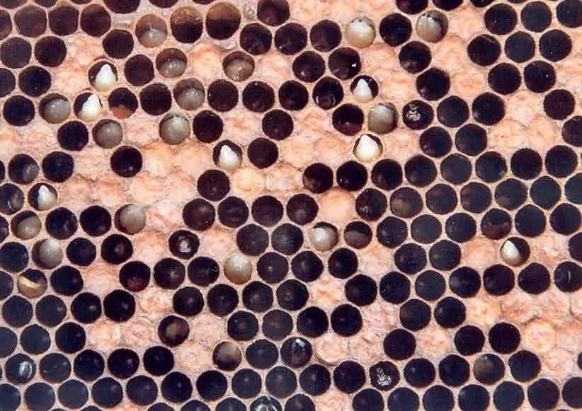
Dead larva remains upright in the cell. Its skin becomes tough and colour changes to pale yellow to brown.
- Head and thorax regions are darker.
- Larva if pulled out with a tweezers, it comes out like a sac.
- Dried scale is boat shaped.
- Sometimes bees desert the colony.
Infection, Multiplication and Spread:
Virus is ingested with food. Two days old larvae are more susceptible. Virus multiplies in body tissues. One dead larva may contain 1 mg virus which is sufficient to infect 1000 colonies. Disease in the colony is spread by nurse bees and among the colonies through swarming, drifting and robbing.
6. Chronic Bee Paralysis
This disease is also called hairless black bee syndrome / little black robbers. Infected bees die within a week. The casual viral particles are irregular in shape. Enzyme ribonuclease found in nectar destroys viral RNA. Hence, there can be recovery with flow of nectar.
Symptoms:
- Bees become hairless, shiny and small.
- Bees have uncoupled wings.
- Affected bees are nibbled by healthy bees, as robbers are nibbled.
- Trembling and jerky movements.
- Sick bees gather on top bars or crawl out.
- Dead bees are seen in front of hive entrance.
7. Iridescent Virus
Symptoms:
- Reduced egg laying / brood rearing.
- Bees become sluggish and make cluster.
- The infected bees crawl on the ground.
- The affected bees cease foraging, reduce honey collection, and the colony face starvation and starts dwindling.
- Illuminated body tissue can be seen under microscope look bluish / greenish.
Spread: Disease is caused by iridovirus. Its infection is serious during hot / dearth seasons. It is transmitted by nurse bees through glandular secretion (food).
Management of Viral Diseases:
- For viral pathogens, there is no chemical control.
- Affected colonies should be isolated beyond their flight range.
- Adopt all the management operations to keep colonies strong.
- Provide proper ventilation to reduce humidity.
- Cage the queen for a week and then requeen.
- Use sterilized equipments / combs.
- Check robbing, drifting and swarming.
- Provide supplement feeding.
- Undertake selective breeding for natural resistance.
II) Adult Bee Diseases:
The diseases of adult bees are caused by protozoa which are single celled animals and form spores or cysts. They multiply by sexual or asexual methods. Their infection reduces vitality of bees, and shortens their life and fecundity. Protozoan is perfect parasites as they do not kill the host immediately. These diseases are difficult to diagnose, though inability to fly, unhooked wings and dysentery can be treated as general symptoms of an unhealthy bee. Microscopic examination is often necessary for a definite diagnosis.
8. Nosema Disease:
This disease is caused by Nosema apis Zander. It is disease of adult bees. It parasitizes all the castes. Their spores germinate in the ventriculus of the host. Pathogen multiplies in epithilial cells and it checks RNA synthesis in the host cells. Its spores are shed in lumen of digestive tract of the affected bee and are then excreted out. One affected bee may contain 180 million spores. Hypopharyngeal glands of the diseased bee are atrophied. Colony strength dwindles down. Infection spreads through ingestion of fecal matter with contaminated food.
Symptoms
- Bees start foraging at younger age.
- Bees feel fatigued, are less able to fly and fall down during their return journey.
- Bees crawl up the grass blades and fall down on the ground and such affected fatigued bees gather in depressions / ditches.
- Abdomen is distended with fecal matter.
- Body hairs are lost and bees become shiny.
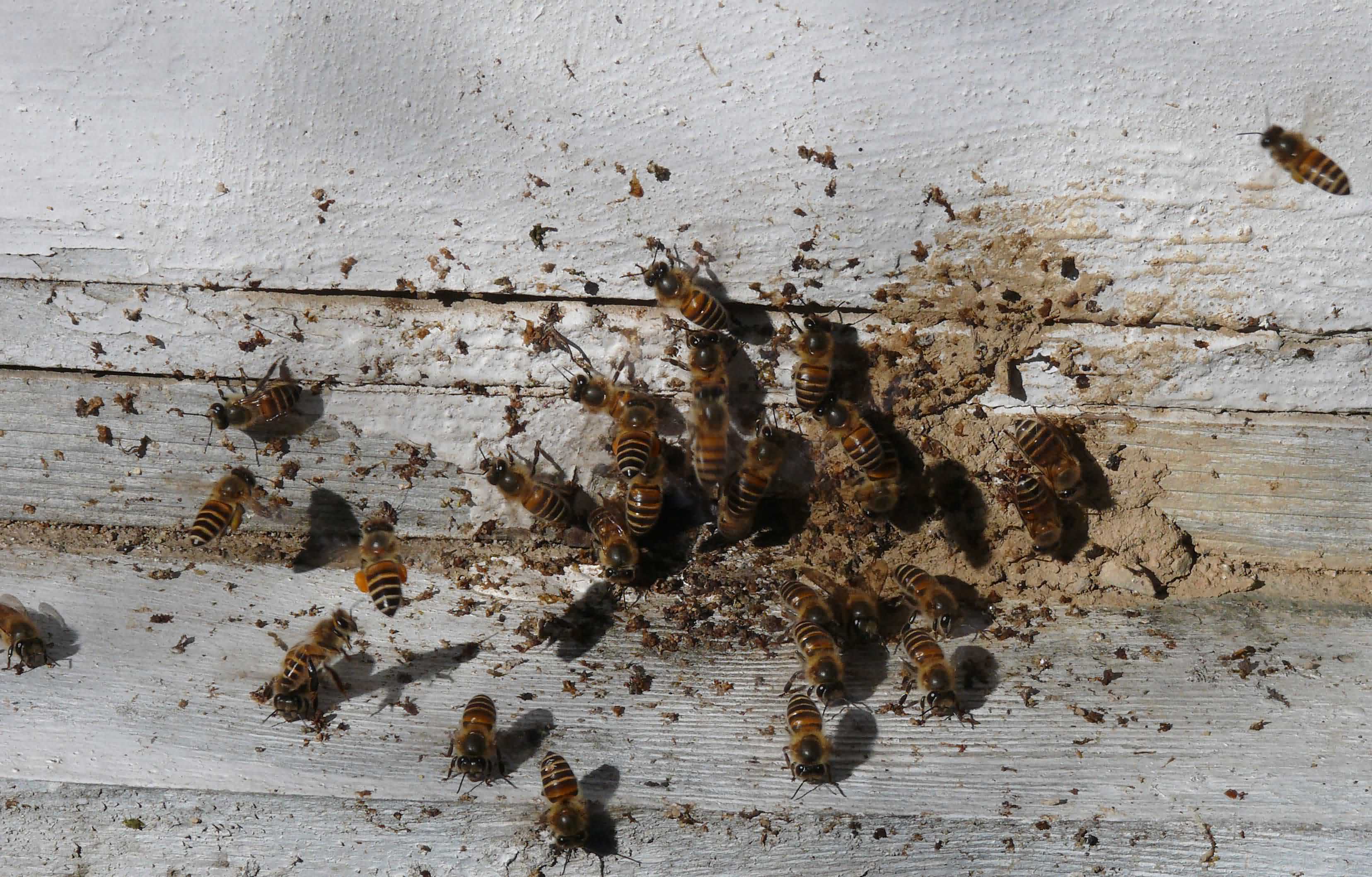
- Mid intestine is swollen and if dissected, shows dull greyish white contents.
- Bees soil the hive entrance (Fig.6 ).
Management:
- Provide fresh running water. Drain off stagnant water from the apiary.
- While transporting queens, select healthy attendant bees.
- Provide upward ventilation to reduce humidity.
- Feed fumagillin in concentrated syrup. It inhibits DNA replication of the pathogen.
- Disinfect the empty hives with ethylene oxide or acetic acid fumigation @ 120 ml / hive.
9. Amoeba Disease of bea
It is caused by Malpighamoeba mellifecae. This infection is caused by ingesting the cysts along with contaminated food.
Cysts germinate, amoeba migrate to malpighian tubes and feed on cell contents. Amoebae multiply by binary fission and form cysts within 18-28 days of ingestion. Cysts accumulate in the mid-gut / rectum.
Cysts are shed in the intestine and are excreted out with the fecal matter. Peak infestation occurs during April-May. Spring dwindling of colony strength can be experienced in such case.
Management:
- Ensure proper hygienic conditions.
- Scarp off the bottom board and disinfect it with 2% carbolic acid.
- Disinfection of hives and equipments with acetic acid is also helpful.
General practices for the management of bee diseases
Honey bees could be affected by diseases and the real cause of abnormality or any disease present in the honey bee broods need to be ascertained before taking up any control measures. It is best to contact the researchers/scientists/beekeeping experts at the nearest centre or university or Government department working on honey bees. After the exact diagnosis of the causal agent of the particular disease, the guidelines/ recommendations given by the expert should be followed in true letter and spirit. However, general advisory for the management of common diseases of honey bees is given below:
(i) Select good site to locate the apiary preferably in an open, dry place with shade.
(ii) Adopt general colony hygiene in the apiary like cleanliness in the beehives including cleaning the bottom board frequently.
(iii) Select and multiply honey bee colonies only from disease resistant stocks.
(iv) Keep colonies with good prolific queens.
(v) Create broodlessness in colony for at least 15 days by enclosing the queen in a queen cage.
(vi) Check the colonies periodically for any abnormalities or changes in behaviour of bees.
(vii)If you observe any colonies with disease, isolate them from healthy ones. Handle diseased and healthy colonies separately.
(viii) Keep the colonies strong by adding sealed brood comb or worker population only from healthy colonies and also by providing adequate food during dearth periods.
(ix) Prevent robbing, drifting, absconding and avoid migration of bee colonies when you notice disease symptoms.
(x) Follow ‘Shook Swarm’ or shaking method to remove contaminated combs completely by transferring entirely new combs in one operation to the colonies with disease symptoms. Destroy the removed combs by burning.
(xi) Sterilise the combs and equipments by any one of the following methods:
- Disinfect the empty combs and equipments with 80 per cent acetic acid @ 150 ml per hive body in piles for few days at a protected place. Air the treated materials before use.
- Dip the contaminated equipments and combs in soap solution containing 7 per cent formalin for 24 hours. Then wash the treated material with water, dry and use.
- Disinfect the combs with UV rays in protected chambers/UV chambers, where possible.
(xii) Use of antibiotics to control honey bee diseases is likely to result in contamination of honey causing problems in export of honey.
Authors:
Sunita Yadav and S. S. Yadav
Department of Entomology
CCS Haryana Agricultural University, Hisar 125 004, Haryana
Mail I D:
