हेलिकोवरपा (HaNPV) के नाभिकीय पॉलीहेडेरोसिस वायरस (एनपीवी) की व्यापक उत्पादन विधि
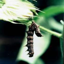
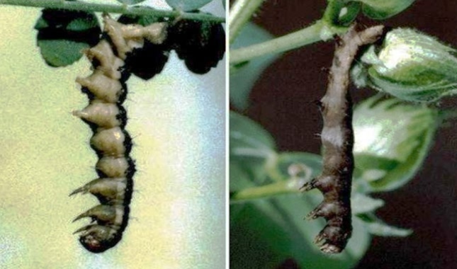
चने और कपास की फसल में एनपीवी से संक्रमित हेलिकोवर्पा आर्मीगेरा का लार्वा
Nuclear Polyhederosis viruses (NPV) are virulent pathogens of insects characterized by the polyhedral occlusion bodies (POB). They are obligate pathogens requiring the host insects for development and multiplication. These viruses are highly specific and do not affect beneficial insects like parasites and predators and are safe to fish, birds, higher animals and man.
Biology of Helicoverpa armigera
Helicoverpa anmigera is the most prevalent, highly phytophagous pest in India and is of most relevance to agricultural economy. It has posed a serious threat to many crops and the apparent importance of the pest calls attention to vital biology of it. Its life cycle on several crops has been studied at several locations by many workers.
Singh.H and Singh.G (1975) have studied the various stages in the lifecycle of Helicoverpa, The larvae pass through six instars. There is vast variation in the colour of the developed larvae. The total larval development period was around 18-20 days.
Effects of H.armigera on various crops
In India the impact of the Helicoverpa annigera on the yield of various aops can differ very widely. Among several aops the importance of H. annigem was seen on the following crops. (Cunningham J.P.et al, 1999).These crops are :
Pigeonpea (Cajanus cajan, Fabaceae), Chickpea (Cicer arietinum, Fabaceae), Maize (Zea mays, Poaceae), Cotton (Gossypiurn sp., Malvaceae), Sorghum (Sorghum bicolor, Poaceae), Sunflowers (Helianthus annnus, Asteraceae), Soybeans (Glycine max, Fabaceae), Tomatoes (Lycopersimm esculentum, Solanaceae), Groundnut (Arachis hypogea, Fabaceae), Alfalfa (Lucerne: Medicngo sativa, Fabaceae), Beans (Phaseolus vulgaris, Fabaceae), Tobacco (Nicotrana tabacum, Solanaceae), and Cucurbits (Cucurbitaceae).
Biology of Nuclear Polyhedrosis Virus (NPV)
The nuclear polyhedrosis virus belong to the family of occluded DNA viruses, Baculoviridae. Representatives of the nuclear polyhedrosis virus (NPV) were the first insect viruses investigated. The virus is characterized by the presence of inclusion bodies, the so-called polyhedra, in infected insects.
The nature and sigruficance of these polyhedra remained a mystery for a long time until the electron microscope was available that the virus particle could be isolated andidentified as the infectious viral agent.
The word, "polyhedra" will designate inclusion/occlusion bodies found in the nucleus of the cells of insects as a result of virus infection. Size and Shape of Polyhedra: The size and shape of polyhedra varies considerably not only between the polyhedra from different insects, but of tenalso within polyhedra of the same species.
Majority of the polyhedral inclusion bodies (PIBs) of H. nmigera NPV are spherical while some of them are irregular in shape. The size ranged from 0.6 pm to 2.3 pm averaging to 1.35 pm. The diameter of polyhedra ranges about 0.5 to 1.5 pm, depending on the insect species. Fig. 6 shows the cross-section of polyhedron of HNPV. In the boundary of polyhedron, the polyhedral envelope (PE) appears as an electron dense layer.
The distance between the polyhedral envelop and polyhedral crystalline matrixis not uniform around the polyhedron. The mean volumc of PIB is 1.29f0.09 pm3 and number of nucleocapsid per polyhedron is 113 f 7.4. The fine structure of polyhedra reveals crystalline lattice of the polyhedra protein molecules, which are arranged in a cubic system.
Although there is no true membrane covering the PIB, difficulties in staining PIB, the retention of their shape, and the presence of a membrane-like coat following chemical and physical treatment indicate that the exterior portion of a PIB is different for the interior portion. On the whole they are very stable and can persist indefinitely in the environment. (Gernot H. Bergold et al, 1982)
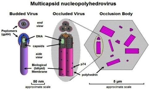
Morphology and size of viruses: Electron microscopic observations are necessary in order to investigate their exact morphology. The viruses can exist in the morphological states free virus particles isolated from polyhedra; virus particles still enclosed in polyhedra and virus particles within infected cell nuclei.
In general they are rod-shaped, about 20-50 nm in diameter (incl. the developmental membrane), and about 200-400 nm long. Virion: Mectious, rod-shaped virions are randomly occluded and singly embedded in PIBs without any apparent disruption of the lattice; an 8 nm layer separates virion from the protein matrix. Alkaline-liberated virions readily lose theu envelopes to reveal nucleocapsids each made up, of a capsid surrounding a DNA core.
The capsid, in turn, consists of protein subunits arranged along its long axis. The virions contain double-stranded, circular DNA molecules present super coiled and packed in the nucleocapsid. Physico-chemical and chemical properties:
It has been known for some time that acids or alkali disrupt PIBs and thus presumably destroy the viral activity. Polyhedra do not dissolve in hot or cold water, alcohol, ether, chloroform, benzol, or acetone.
The Viruses
The virus consists of protenaceous polyhedral occlusion bodies inside which the virions or rod shaped viruses are embedded. The virions are made up of DNA and belong to the Baculovirus group.


Mode of entry and infection
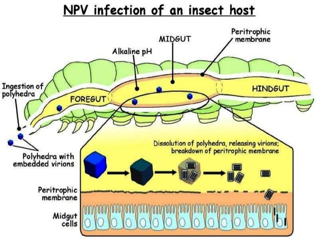
To produce the disease, the virus should be ingested. This is otherwise called as per as infection. Soon after entry into the insect gut, due to the action of the alkaline gut juice followed by the action of proteolytic enzymes, the polyhedral coat is loosened and dissolved resulting in the liberation of the virions.
The liberated virions pass through the mid gut cells of different tissues like fat body, tracheal matrix, haemocytes, sarcolemma of muscles, neurilemma and nerve cells of ganglion and brain, hypodermis and gonads.
Symptoms of the diseases:
 Infected insect become dull in color and inactive. Feeding rate is very much reduced and color of larvae. turns pale and as the disease advances, Larvae develop pinkish white coloration especially in the ventral aspect due to the accumulation of the polyhedral.
In advanced stages of infection the larvae become flaccid, the skin becoming very fragile and rupturing on even slight disturbance. Whitish fluid containing thousands of POB exudes from the ruptured skin.
Some of the diseased larvae can be found hanging head downward from the plants or from the lid.
Material required:
Helicoverpas armigera NPV Materials required
- Empty injction vials
- Absorbant cotton
- Soaked Bengal gram seeds.
- Semisynthetic diet
- NPV suspension
- Fourth instar larvae of armigera (Fourth instars larvae will have a mean head capsule width of 1.45 mm and will reach this stage within 5-7 days).
- Equipments- as indicated under litura.
Ingredients of Semi-synthetic diets:
|
Sr.No. |
Ingredients |
Quantity |
|
(A)1. |
Agar Agar |
12.75 grams |
|
2. |
Distilled Water |
390 ml |
|
(B)1. |
Gram flour |
105 grams |
|
2. |
Methyl Parahydroxy Benzoate |
2 gram |
|
3. |
Yeast powder |
10 grams |
|
4. |
Ascorbic Acid |
3.5 gram |
|
5. |
Sorbic Acid |
1 gram |
|
6. |
Streptomycin |
0.25 gram |
|
7. |
Multivitamin |
1Cap |
|
8. |
Distilled water |
390 ml |
Boil Part A portion separate and added into part B and pour into Test tubes.
Production methodology of virus
- Starve the fourth instars larvae for 8 hrs.
- Dip the heads of these larvae in the NPV suspension containing about 108 POBS/ml.
- Rear in individuals vials on soaked Bengal gram seeds. The seeds should be changed daily after removing the excreta.
- Virus inoculated larvae can also be reared until death on semi synthetic diet with all ingredients except formalin. This can save the labour of changing be larval food daily.
- Larvae will begin to show symptoms of infection in about 4 days and begin to die by 5-6 days.
- Collect larvae which have died, (exhibiting the characteristics symptoms) in distilled water.
- Purify as described already.
- Virus obtain from 750 virosed larvae will yield approximately 1.5 multiplied by 1012 POB will be sufficient to treat on hectare.
Albistriga NPV
Fourth instar larvae (2.5-3cm) are ideal for mass culturing of the virus with an inoculums dose of 108 POB/ml for maximum harvest of NPV.
The materials required are more or less the same as in S. litura except that instead of castor leaves, Calotropis leaves are used.
Steps in the culturing of virus
- Ten day old (4th instar) larvae measuring 2.5 to 3 cm are ideal for he culturing of NPV.
- Collect them and starve overnight.
- Prepare virus suspension in 250 ml water with few drops of teepol (containing 108 POB/ml).
- Treat Calotropis leaves after removing the wax coating with cotton wool thoroughly with the virus suspension and dry in shades for few minutes.
- Provide treated leaves to the starved caterpillars to feed.
- On the second day also provide the treated leaves; subsequently untreated leaves are given as food.
- Larvae with typical symptoms will start dying from 5-6 days of inoculation. (In infected larvae the skin on the under surface turns pinkish white due to the accumulation of polyhedra). After death, the skin ruptures and white liquefied body contents oozes out.
- Collect the dead larvae and filter through clean muslin cloth. Allow the virus to settle in sufficient water.
- After a week, pour out the supernatant alone carefully, add water and allow it to settle again.
- The sediment (white in color) is taken and purified as indicted already.
Polyhedral obtained from 500 virosed (big sized) larvae (1.5 multiplied by 1012 POB/AC) will be sufficient for an acre.
Precautions
- Brackish water for handling virus.
- Grown up caterpillars for inoculation.
Authors
Dr T.A Usmani & Rajeev Kumar
Regional Central Integrated Pest Management Centre,
Sector-E, Ring Road, Jankipuram, Lucknow,Uttar Pradesh
Email:

